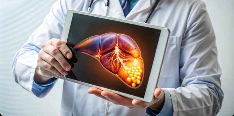
A liver tumour may be discovered during a routine health check-up, ultrasound, or investigation of symptoms such as abdominal pain, unexplained weight loss, loss of appetite, or jaundice. Finding a tumour does not always mean that it is cancerous. Some liver tumours are benign, while others may require urgent medical attention. Accurate diagnosis helps doctors understand the type, size, location, and extent of a tumour. For people seeking Liver Tumour Treatment in Delhi Ncr, appropriate imaging and specialist evaluation are important before deciding on treatment. Dr. Pulkit Rangarh focuses on detailed radiological assessment to help evaluate liver lesions and support treatment planning.
Want a direct connection with us on WhatsApp? Click now.
What Is a Liver Tumour?
A liver tumour is an abnormal growth of cells in the liver. It can be benign or malignant.
Benign tumours are non-cancerous and may not require immediate treatment. Some remain stable and only need regular monitoring. However, larger lesions can sometimes cause abdominal discomfort or pressure on nearby organs.
Malignant tumours are cancerous and require timely evaluation. Cancer can begin in the liver itself, known as primary liver cancer. It can also spread to the liver from another part of the body. This is called a metastatic or secondary liver tumour.
Because different liver lesions can have similar appearances, proper diagnostic testing is important for planning Liver Tumour Treatment in Delhi Ncr.
How Is a Liver Tumour Initially Detected?
Diagnosis usually starts with a medical history and physical examination. The doctor may ask about symptoms, previous liver problems, hepatitis infection, medications, and other relevant health conditions.
Blood tests may also be recommended. Liver function tests can provide information about liver health. In selected cases, doctors may check tumour markers such as alpha-fetoprotein (AFP).
However, blood tests alone generally cannot confirm the exact nature of a liver tumour. Imaging tests are therefore an important part of the diagnostic process.
What Is the Role of Ultrasound?
Ultrasound is often one of the first imaging tests used when a liver abnormality is suspected. It uses sound waves to create images of the liver and does not involve radiation.
An ultrasound can detect a liver mass and provide information about its size and general appearance. It can also help identify other abnormalities in the abdomen.
If the ultrasound shows a suspicious lesion or does not provide enough information, the doctor may recommend a CT scan or MRI for a more detailed assessment.
When Is a CT Scan Recommended?
A CT scan uses X-rays and computer technology to create detailed cross-sectional images of the liver and surrounding structures. A contrast-enhanced CT can provide additional information about the blood supply and appearance of a liver lesion.
CT imaging can help doctors assess:
- The size and exact location of the tumour
- The number of liver lesions
- Involvement of nearby blood vessels
- The relationship with surrounding organs
- Possible spread of the disease
CT can be particularly useful when doctors need a broader assessment of the abdomen and chest. It can also provide valuable information for treatment planning.
For patients undergoing evaluation for Liver Tumour Treatment in Delhi Ncr, CT findings may help specialists determine the stage and possible treatment approach.
When Is an MRI Recommended?
MRI uses magnetic fields and radio waves to create detailed images of soft tissues. Unlike CT, it does not use ionising radiation.
A liver MRI can provide excellent detail about the internal structure and characteristics of a lesion. It may be recommended when a CT scan does not provide sufficient information or when doctors need to examine a tumour more closely.
Contrast-enhanced MRI can show how a lesion behaves during different phases of blood flow. This can help doctors distinguish between certain types of liver lesions.
MRI can be especially helpful for complex tumours, multiple lesions, or abnormalities located near important blood vessels. Detailed imaging can support more personalised Liver Tumour Treatment in Delhi Ncr.
Is a Biopsy Always Necessary?
No. A biopsy is not required for every liver tumour.
During a biopsy, a small sample of tissue is collected from the lesion and examined under a microscope. This can help determine whether abnormal cells are cancerous and identify the type of tumour.
However, some liver cancers have characteristic appearances on high-quality CT or MRI. In such situations, imaging may provide enough information for diagnosis without a biopsy.
A biopsy may be considered when imaging findings are unclear, when tissue confirmation is needed, or when the result could affect the treatment decision.
The decision depends on the patient’s medical history, imaging findings, suspected diagnosis, and overall treatment plan.
How Do Doctors Choose the Right Test?
There is no single diagnostic test suitable for every liver tumour. Doctors consider the patient’s symptoms, medical history, liver condition, lesion size, imaging appearance, and previous test results.
A diagnostic process may begin with an ultrasound. If a suspicious lesion is found, contrast-enhanced CT or MRI may be advised. A biopsy is considered only when it is medically appropriate.
This step-by-step approach helps doctors gather important information while avoiding unnecessary investigations.
Why Is Early Diagnosis Important?
Early diagnosis can make an important difference in liver tumour management. Some benign tumours may only require observation, while malignant tumours may need active treatment.
Depending on the type and stage, treatment options may include surgery, ablation, embolisation procedures, systemic medicines, or a combination of approaches.
Imaging also helps doctors understand the tumour’s relationship with blood vessels and nearby organs. This information can influence which treatment is suitable.
Therefore, patients searching for Liver Tumour Treatment in Delhi Ncr should have their liver lesion properly evaluated before treatment begins.
Conclusion
Diagnosing a liver tumour requires careful assessment rather than relying on a single test. Ultrasound may provide the initial evaluation, while CT and MRI offer more detailed information about the tumour’s size, location, characteristics, and possible spread. A biopsy is recommended selectively when tissue confirmation is necessary.
For patients looking for Liver Tumour Treatment in Delhi Ncr, accurate diagnosis is an essential first step toward choosing an appropriate treatment strategy. Timely specialist evaluation and suitable imaging can help patients understand their condition and move forward with an informed treatment plan.
For more information or to schedule a consultation, visit https://share.google/bI1IzCyOg3uUQW9n3 and take the first step toward achieving your dream.